46
SKIN CARE TODAY
2015,Vol 1, No 1
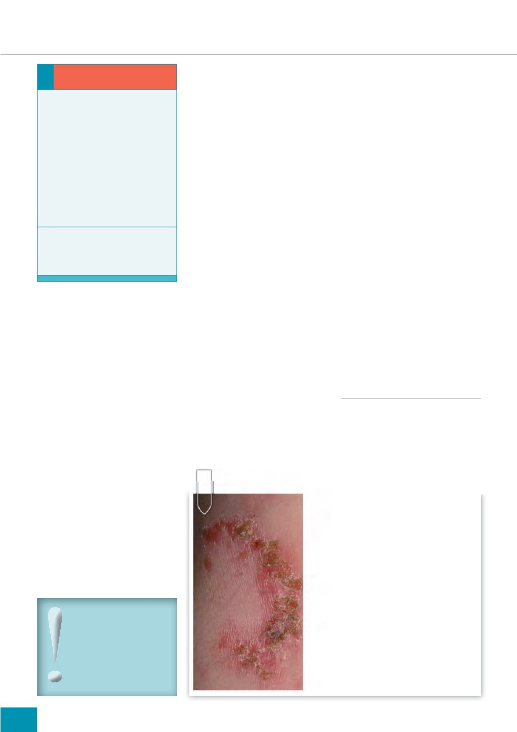
into gold-crusted plaques, typically
2cm in diameter (these have been
described as resembling glued-on
cornflakes). Satellite lesions may
also occur due to autoinoculation
(self-infection) (NICE, 2013a).
Bullous impetigo is characterised
by flaccid, fluid-filled vesicles and
blisters (bullae), between 1–2cm
in diameter. When these rupture
they leave the skin raw and form
thin, flat, brown-to-golden crusts.
The lesions are multiple and spread
rapidly. They are also painful and
the patient may develop systemic
symptoms (weakness, fever and
diarrhoea), and lymphadenopathy
(swelling of the lymph nodes).
What tests should be done?
Skin swabs are not necessary to
diagnose impetigo. Instead, swabs
should only be used to identify the
bacteria involved and its sensitivity
to antibiotics if the infection is
(NICE, 2013a):
i
Very extensive or severe
i
Recurrent, in which case a nasal
swab for Staphylococcal carriage
could be considered (nasal
carriage of
S. aureus
is a known
risk factor for skin infections)
i
Suspected as being a
community outbreak
i
Suspected as being caused by
meticillin-resistant
S. aureus
(MRSA).
How is impetigo treated?
Localised non-bullous impetigo
should be treated with topical
fusidic acid (three to four times
daily, for seven days [eMC, 2013]).
Before it is applied, the crusts of
any plaques should be removed
by soaking them in soapy water
(as long as this does not cause
discomfort). Removal of the crust
allows the antibiotic to come into
direct contact with the bacteria
rather than being wasted on dry,
exfoliating skin (Watkins, 2005).
Topical antibiotics (mupirocin
and retapamulin) are not
recommended as a first-choice
treatment; neither are topical
antiseptics, as there is a lack of
evidence to support their efficacy
(Koning et al, 2012).
If the impetigo is bullous,
extensive, or severe with
systemic symptoms, oral
antibiotics are the first-choice
treatment (NICE, 2013a).
Complications
The infection may spread locally
and systemically, resulting
in cellulitis (infection of the
deeper layers of the skin and the
underlying tissue), lymphangitis
(inflammation of the lymphatic
system), or septicaemia (invasion of
bacteria into the bloodstream).
Non-infectious complications
of
S. pyogenes
infection include
guttate psoriasis (an acute
skin eruption), scarlet fever,
and glomerulonephritis (an
inflammation of the kidney that can
lead to kidney failure) (Koning et al,
2012).
More rarely, exotoxins (toxins
secreted by bacteria) produced
by some strains of
S. aureus
may
result in staphylococcal toxic shock
syndrome or staphylococcal scalded
skin syndrome (SSSS) (results in
widespread formation of fluid filled
blisters) (DermNet NZ, 2013a).
FUNGAL INFECTIONS
What are fungal infections?
Fungal infections of the skin (tinea)
are caused by dermatophytes or
fungi that require keratin for growth
— keratin being the key structural
Did you know:
%XOORXV LPSHWLJR OHVV
FRPPRQO\ DIIHFWV WKH IDFH
PRUH RIWHQ GHYHORSLQJ RQ
WKH D[LOOD XQGHUDUP QHFN
IROGV DQG ¶QDSS\· DUHD
1,&( D
FOCUS ON SKIN INFECTIONS/INFESTATIONS
i
THE SCIENCE — HOW DOES THE SKIN
BECOME INFECTED?
Skin infections such as impetigo occur when
bacteria (such as
Staphylococcus
) access a
break in the skin, such as a cut or crack in
dry skin. This results in symptoms such as
boils or abscesses — pus-filled lumps on
the surface or just under the skin, which are
often painful. This in turn can lead to a crust
on the skin (impetigo), or redness, swelling
and pain in the underlying tissue (cellulitis).
If these conditions are not treated, invasive
infections can develop, which have more
severe and wide-ranging symptoms
including fever, low blood pressure,
confusion and shortness of breath.
Source:
Credit: Evanherk at nl.wikipedia
Impetigo — facts...
1RQ EXOORXV LPSHWLJR
(impetigo contagiosa or
crusted impetigo) is the most
common form, accounting for
three-quarters of cases, with
Staphylococcus aureus
being
the main cause.
Streptococcus
pyogenes
is implicated in fewer
cases, which develop either
due to
S. pyogenes
alone or in
combination with
S.aureus
and
bullous impetigo (NICE, 2013a).
%XOORXV LPSHWLJR
is always
caused by
S. aureus
(Cole and
Gazewood, 2007; Koning et al,
2012).


